CT Scans of Egyptian Priests Nes-Min & Nes-Hor Reveal Chronic Back Pain, Dental Disease, and the Realities of Ancient Life.lh
CT Scans of Egyptian Priests Nes-Min & Nes-Hor Reveal Chronic Back Pain, Dental Disease, and the Realities of Ancient Life
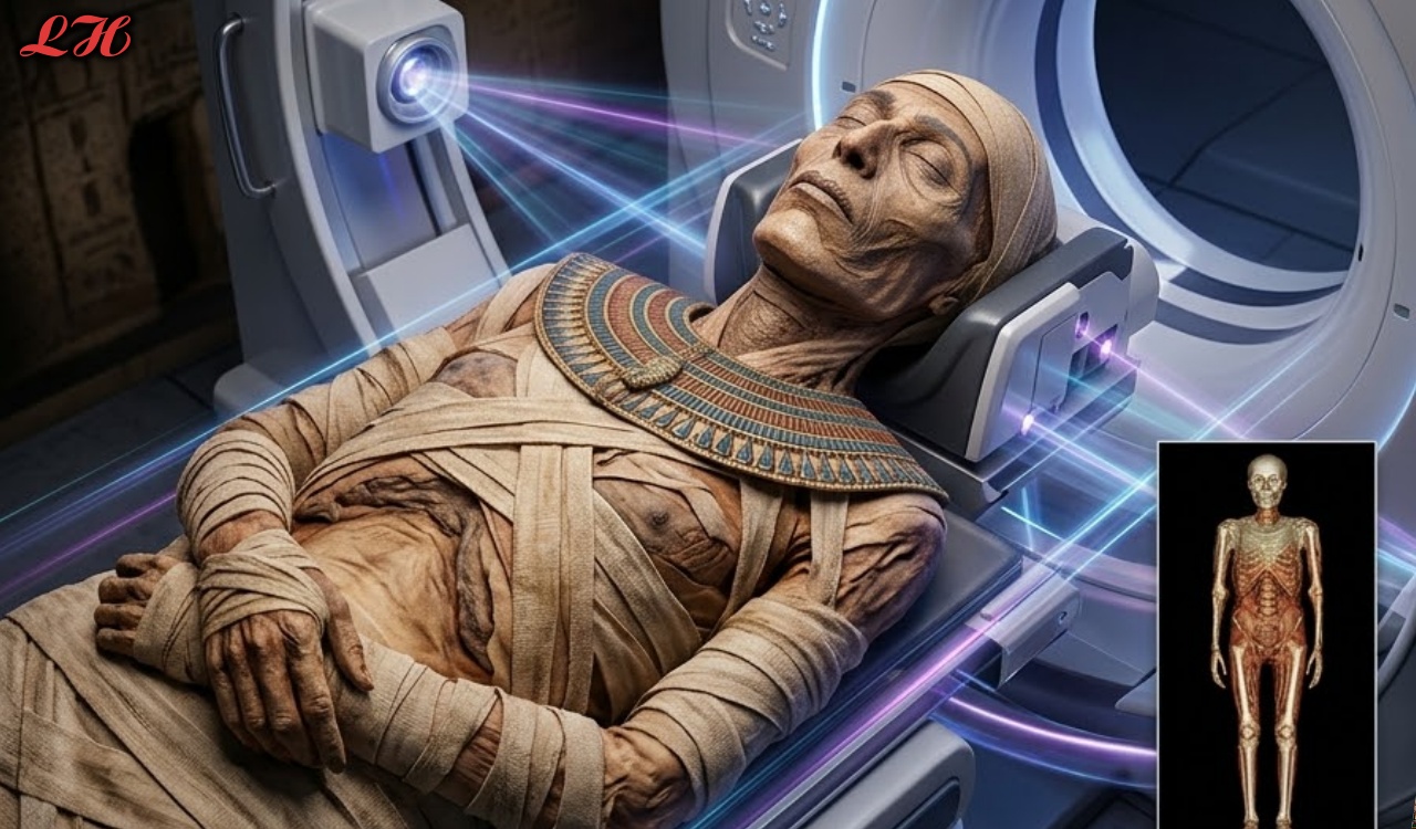
In a February 2026 study from Keck Medicine of USC, high-resolution 320-slice CT scans of two Ptolemaic-era Egyptian priest mummies have delivered an unprecedented medical portrait of daily life more than 2,200 years ago. Nes-Min (died ~330 BCE in his forties) and Nes-Hor (died ~190 BCE in his sixties) both suffered from familiar modern ailments—chronic pain, dental decay, and joint degeneration—proving that even temple elites were not immune to the physical toll of aging and labor.
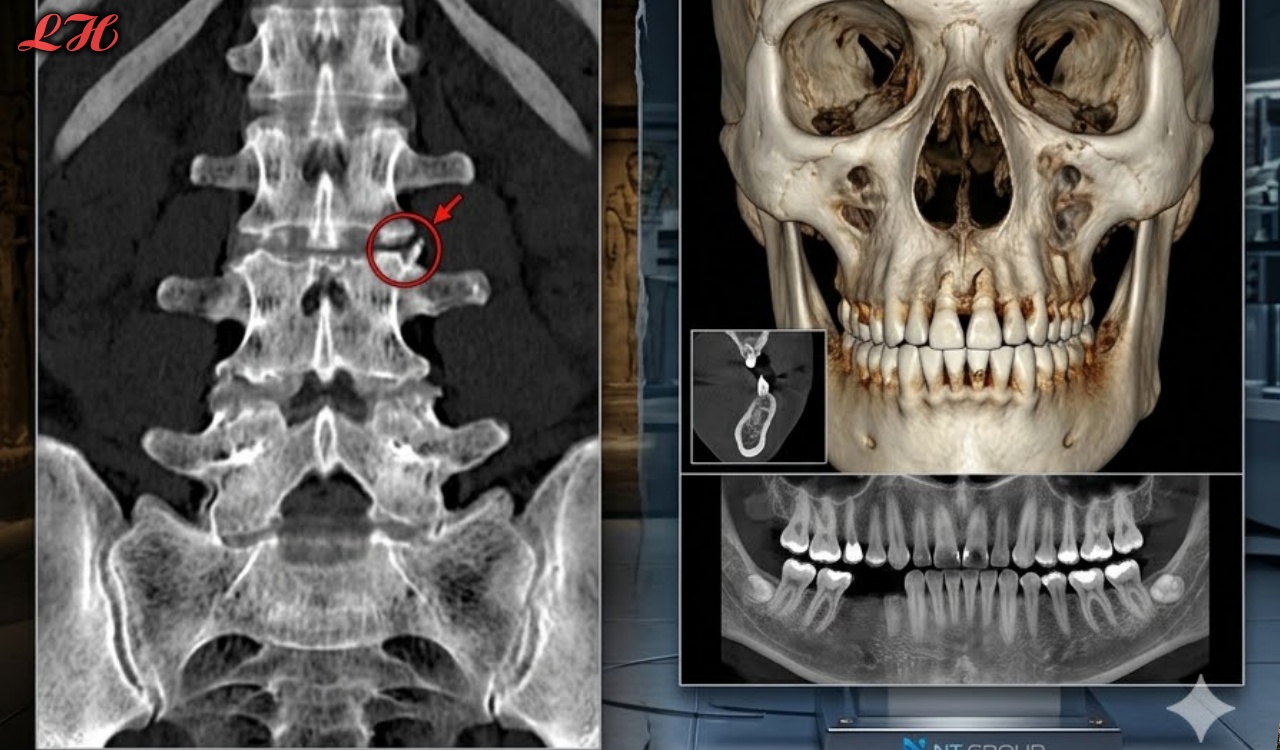
Scanned while still inside their heavy sarcophagi, the mummies revealed intimate details impossible to see through traditional unwrapping. Nes-Min’s lumbar spine showed a collapsed vertebra consistent with long-term strain and age-related wear, mirroring the chronic lower-back pain experienced by millions today. Nes-Hor, who lived longer, endured advanced dental disease and a severely deteriorated hip joint that would have caused visible limping and mobility loss.
“These scans humanize them,” said radiologist Summer Decker. “We see eyelids, lips, healed injuries, and the exact pathologies that caused real suffering.” Burial goods—scarab beetles, a fish, beaded necklaces, and net garments—were also mapped without disturbance, adding cultural context.
The findings underscore that ancient Egyptian priests faced the same degenerative conditions as contemporary humans, despite advanced mummification. By merging cutting-edge medical imaging with archaeology, the project transforms two anonymous mummies into individuals whose bodies tell stories of resilience, hardship, and the universal human experience across millennia.