Modern Medicine Meets Ancient Mystery: CT Scans Reveal the Secret Lives of Egyptian Priests

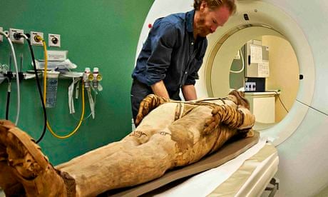
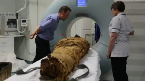
In a remarkable fusion of 21st-century technology and ancient history, radiologists at Keck Medicine of USC have utilized state-of-the-art 320-slice CT scanning to peer through the linen wrappings of two Egyptian mummies. The results, released in early 2026, have provided an unprecedented look at the health, physical struggles, and daily lives of two priests who lived over 2,000 years ago.
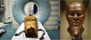
The project focused on Nes Min (circa 330 BCE) and Nes Hor (circa 190 BCE). In a technically challenging feat, each mummy was scanned while still resting in the lower half of its original, 200-pound stone sarcophagus. The high-resolution imaging allowed researchers to “unwrap” the priests digitally, revealing physical details so fine that even eyelids and lower lips were visible on the 3D models.
Echoes of Modern Ailments
What surprised researchers most was how closely the physical burdens of these ancient men mirror the health issues of people today. The scans revealed that even the priesthood was not immune to the wear and tear of time:
-
Nes Min (The Younger Priest): Though he lived during the transition to the Ptolemaic period, Nes Min’s spine tells a story of chronic discomfort. Scans showed a collapsed lumbar vertebra, a condition typically associated with long-term strain and aging. He likely suffered from persistent lower back pain throughout his career.
-
Nes Hor (The Elder Priest): Despite living in a later era, Nes Hor reached a more advanced age but faced greater physical challenges. His results revealed severe dental decay and a deteriorated hip joint. Experts believe his hip condition was so advanced that it likely caused a significant disability, making walking painful and difficult.

Hidden Treasures and 3D Recreations
Beyond pathology, the scans uncovered “hidden” burial traditions. Within the wrappings of Nes Min, radiologists identified several small artifacts that had remained unseen for millennia, including symbolic scarab beetles and a fish-shaped amulet.
To bring these findings to the public, the team at the USC Center for Innovation in Medical Visualization used medical-grade 3D printers to create life-size replicas of the priests’ skulls, spines, and hips, as well as the buried artifacts. These touchable pieces of history, along with the digital models, are currently featured in the “Mummies of the World” exhibition at the California Science Center.
A New Window into History
“These mummies were scanned in the 1990s, but today’s technology offers a much higher resolution,” explained Dr. Summer Decker, director of the Center for Innovation in Medical Visualization. By applying the same tools used to plan complex surgeries for modern patients, the team has successfully humanized these ancient figures.
The study serves as a powerful reminder that while empires rise and fall, the human experience—from the artistry of a beaded burial shroud to the simple ache of an aging back—remains a constant across the ages.